MediVisuals

Scroll down to view blog archive

Scroll down to view blog archive

Wednesday, August 6, 2014

Discogenic Pain - Back Pain and Radiculopathy Without Evidence of Significant Disc Injury

 By: Robert Shepherd MS, Certified Medical Illustrator, President & CEO, MediVisuals Inc.

In determining if a person's pain may be related to some sort of intervertebral disc pathology, a great deal of emphasis is placed upon imaging studies showing evidence of mechanical compression of a nerve root by abnormal posterior displacement of a disc (i.e. bulge, protrusion, herniation, etc.) as portrayed in the below illustration.


In cases where clear mechanical compression of the nerve roots is not shown in imaging studies, some are quick to argue that any pain emanating from the area is either exaggerated or entirely contrived. However, a person can experience pain consistent with mechanical compression of a nerve root without having any significant disc pathology. This is because the spine is encircled with a meshwork of nerves that are much too small to be seen on CT or MRI (see the below figure). The sinuvertebral nerves surround and penetrate the intervertebral discs.
When injuries to a disc are more subtle, the sinuvertebral nerves may detect the injuries and send pain signals to the brain where they are interpreted as pain (see figure). The pain may be limited to the area of the back, or a pain perception phenomenon know as "pain referral" (confusion of the origin of pain signals by the brain) may result in the person experiencing very real pain consistent with radicular pain from mechanical nerve root compression by a severely herniated disk.
Another common cause of pain consistent with nerve root mechanical compression is chemical irritation or inflammation of the nerve root. Chemical irritation of a nerve root often results from the release of chemicals following a more subtle disc injury (see the below figure). These chemicals irritate and inflame the nerve root and surrounding tissues, resulting in the perception of pain consistent with an injury to the disc and mechanical compression of the nerve root. Even after resolution of chemical irritation and inflammation, scar tissue may develop that binds the nerve root (often undetectable on CT or MRI). This scarring can cause permanent debilitating pain that may require surgical intervention.

© MediVisuals, Inc. - Permission to use any image (or parts thereof) posted on this blog in depositions, demand packages, settlement hearings, mediation, trial, and/or any other litigation or non-litigation use can be obtained by contacting MediVisuals at trose@medivisuals.com – otherwise copyright laws prohibit their use for those or other purposes. 

Friday, May 16, 2014

Disc Herniation and Other Disc Injuries

By: Robert Shepherd MS, Certified Medical Illustrator, President & CEO, MediVisuals Inc.

The term “degenerated disc” is generally used to describe a disc in the early degenerative process. It is the beginning of a progressive break down of the disc. This condition can be initiated or accelerated by a traumatic event.


A disc bulge is a more advanced collapse of the disc to the point that the disc expands beyond its normal contour. It may or may not impinge on the neural structures within the spinal canal or neural foramina. Similarly, a disc bulge can be the immediate or delayed result of a traumatic event, or a traumatic event may exacerbate a preexisting, stable disc bulge.


A subligamentous herniation is one in which nucleus pulposus has extended through the annulus fibrosus, but has not gone through the posterior longitudinal ligament.


The term “herniation” is generally used when the nucleus has completely extruded through the annulus fibrosus and posterior longitudinal ligament

However, it does not matter what the disc pathology is labeled, if it impinges upon, or irritates the neural components, it is a significant injury that will likely require some type of invasive procedure to correct. 

The disc can either be injured by an immediate tear of the annulus fibrosus and extrusion of the nucleus pulposus during a traumatic event, or they can be the result of a much more gradual process. In order to understand the gradual breakdown of the disc, one must first understand a little of the physiology of a disc. The inner disc relies on exchange of fluid, nutrients and oxygen through the end plate of the adjacent vertebral bodies.

During a traumatic event, the endplate may become injured resulting in interference with that exchange.
As a result of the inability of the disc to obtain the fluid, nutrients, and oxygen it needs, the disc gradually begins to break down - becoming a degenerated disc, followed by a bulging disc, and eventually to a herniated disc. The amount of time involved with the process of the disc breakdown is related to the severity of the initial disc injury.


Therefore, if a disc injury is not evident until weeks or months after a traumatic event, it does not mean the injury was not a direct result of the traumatic event.


© MediVisuals, Inc. - Permission to use any image (or parts thereof) posted on this blog in depositions, demand packages, settlement hearings, mediation, trial, and/or any other litigation or non-litigation use can be obtained by contacting MediVisuals at www.medivisuals.com – otherwise copyright laws prohibit their use for those or other purposes.

Wednesday, May 14, 2014

Breaking Down Traumatic Arthritis

 By: Robert Shepherd MS, Certified Medical Illustrator, President & CEO, MediVisuals Inc.

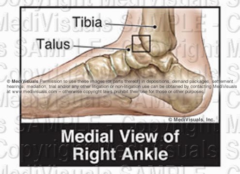
Fractures can result in several long term or permanent complications that can necessitate additional surgical procedures. One of the most common long term debilitating complications is traumatic arthritis.Traumatic arthritis can affect almost any moveable joint in the body. To explain traumatic arthritis more in depth, we will be focusing on the tibiotalar (ankle) joint, as shown in the illustration below.


 Buy on PI Exhibits NOW

As like most moveable joints, the tibiotalar joint consists of smooth articular bone covered by thick, shock-absorbing articular cartilage. 


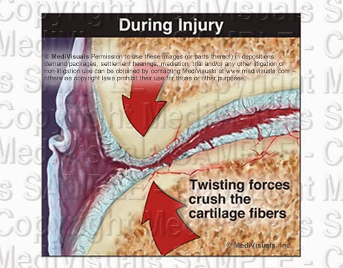
During trauma, the joint surfaces can be driven together resulting in injuries to the cartilage and microfractures of the articular surface (even without obvious intrarticular fracture). 


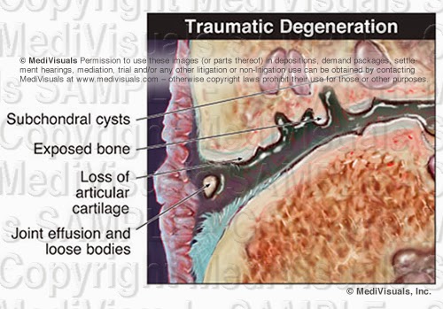
The bone and cartilage then undergo changes that result in the progressive breakdown of the joint. As the process advances, the joint becomes painful. In most cases, the only treatment options are joint replacement or fusion.


The acromioclavicular (AC) joint also frequently falls victim to traumatic arthritis. As the AC joint enlarges (hypertrophy) it impinges on the rotator cuff, which is referred to as subacromial impingement. This can cause irritation or tearing of the rotator cuff and is most often treated by AC joint resection and subacromial decompression.


Tuesday, April 22, 2014

Cervical Connective Tissue Injury (a.k.a. “Whiplash,” “Cervical Soft Tissue Injury,” and “Cervical Strain and/or Sprain”)

By: Robert Shepherd MS, Certified Medical Illustrator, President & CEO, MediVisuals Inc.

"Cervical Connective Tissue Injury," "Cervical Soft Tissue Injury," "Cervical Strain," and "Cervical Sprain" are among the common names used to label specific injuries to the muscles, tendons, and ligaments of the cervical spine resulting from trauma. Proving the presence of such injuries in litigation is particularly challenging for a number of reasons. First, there is no imaging study or medical test that can definitively reveal these types of injuries as they typically exist, with the exception of an invasive biopsy of the affected tissues. Health care providers typically make the diagnosis based upon a number of factors--primarily upon the appearance of symptoms soon after a related traumatic event as well as at least partial relief of symptoms following specialized therapies.

The second factor that greatly contributes to the difficulty of these cases is that, in years past, these same injuries were commonly referred to as "whiplash". It would be difficult to find an individual (juror, mediator, judge, or arbitrator) who does not have preconceived negative connotations associated with the term "whiplash." Almost certainly, those defending claims involved with the existence of the condition will dismiss any other label of the condition except "whiplash" when arguing a case.

To overcome the hurdles encountered by those arguing that their plaintiffs do indeed suffer from these injuries, it is important to combat the voodoo and skepticism associated with the term "whiplash" and explain the well-founded science associated with the injuries and how they result from the trauma of a particular event. Illustrations explaining the related anatomy, the mechanism of injury, and the injury itself can be very valuable tools in that process. Following are examples of graphics that have been successfully used many times for those purposes.

These particular illustrations and the concepts behind them were developed by MediVisuals in 1996 after extensive research into the anatomy, the mechanism of injury, and the resulting anatomical trauma which occurs in these cases.


Cervical Connective Tissue Injury

The above group of images is helpful in explaining the general mechanism of injury to the cervical muscles, tendons, and ligaments during a typical automotive sudden deceleration or acceleration event. The large illustration of the upper torso, neck, and head in extreme forward flexion (hyperflexion) shows many of the posterior muscles of the neck that are subject to cervical strain injuries and their bony attachments. The greater occipital nerve is included as it is sometimes involved and can result in post-traumatic occipital headaches.

A diagram in the upper right corner demonstrates hyperextension and hyperflexion. The image also demonstrates some of the major ligaments of the neck which may also be injured as a result of these violent and extreme motions. These ligaments may be injured grossly or microscopically by excessive flexion, extension, or rotation.

Three illustrations in the lower right show a close-up view of the spinal anatomy 1) in the Normal condition, 2) during Excessive Stretching, and 3) After Healing. In the normal view, the close relationship between the muscles, nerves, and blood vessels is pictured and can be appreciated. Shown in the illustration depicting excessive stretch are small tears and the resulting bleeding in the muscle fibers. In the final illustration, the relationship of the structures after healing is depicted. Scar tissue and inflammation entrap blood vessels and nerves, resulting in a permanent state of compromised, painful movement.


Microscopic Appearance of Muscular and Tendon Strain Injury

The above series of illustrations compares the Normal relationships of structures to those observed during Excessive Stretch and After Healing. The series can be used to explain excessive stretch injuries in any area of the body (neck, back, temporomandibular joint, etc.). Each illustration depicts the structures at sequentially higher magnifications, beginning with the largest center image in each category.

The Normal series of illustrations begins by depicting nerves and small blood vessels intertwined in muscle and its tendinous attachment to the bone. The second illustration in the group shows a magnified view of a muscle fiber. Complicated and highly organized relationships of the many delicate structures necessary for proper function are depicted. The third illustration at the top of the panel details the relationships of the myofilaments (actin and myosin) in their normal relaxed position.

The Excessive Stretch series of illustrations begins by depicting the muscle, tendon, blood vessels and nerves as they are excessively stretched. Small hemorrhages are seen escaping from the stretched and torn blood vessels. In the second illustration, blood is shown escaping into surrounding spaces, reducing oxygen exchange to the muscle and irritating the delicate structures of the muscle fiber. The uppermost illustration, depicting the myofilaments, shows the actin and myosin fibers torn and stretched past the point of normal interdigitation.

The last series of illustrations depicts the permanent injuries that result After Healing. The center illustration shows scar tissue and inflammation occluding blood vessels and adhering the delicate structures of the muscle fibers together, limiting motion and causing chronic pain and muscle spasm. The bottom illustration shows the irregular outline of the scarred and inflamed muscle fiber. Small adhesions are seen between the blood vessels, nerves, and muscle fibers. The uppermost illustration shows the damaged myofilaments. Their normally well-organized interdigitating arrangement is destroyed, limiting muscle movement at the most basic level. 

Collectively, all these injuries prevent normal free and painless movement. Instead, the scarring binds the structures together, entrapping capillaries that results in reduction in blood supply. Nerves are also entrapped, resulting in pain. Lastly, during movement the binding of structures results in irritation, inflammation, and muscle spasm.

Citations available upon request.


Wednesday, April 9, 2014

Complications Associated with Fracture Injuries

 By: Robert Shepherd MS, Certified Medical Illustrator, President & CEO, MediVisuals Inc.

Several long term or permanent complications may result from a fracture injury. Traumatic arthritis may be one of the most common long term debilitating complications. (See the previously posted article "Breaking Down Traumatic Arthritis" for more information.) These complications may even require additional surgical procedures.

Normally, fractures begin healing by forming a callus which is then replaced by bone. With a nonunion (see image below), the callus is replaced by fibrous tissue instead of by bone. This is a painful condition that is most frequently treated by surgery to remove the fibrous tissue, debride the surrounding bone and re-approximate the remaining fragments (sometimes with bone graft material and fixation devices).


Malunion is another frequently occurring complication of fractures. With malunion, the bones heal, but they do so at an unusual angle that can effect range of motion and/or cause pain (see image below). Treatment typically involves refracturing or cutting the bones, realignment, and fixating them with hardware so that they heal in the correct position.


Intra-articular “step-offs” are a complication of intra-articular fractures. A “step-off” is a type of malunion in which the bone heals, but it heals so that the joint surfaces are not aligned in a smooth continuous surface. The illustration below shows a fracture through the patella that healed with a painful “step-off.”


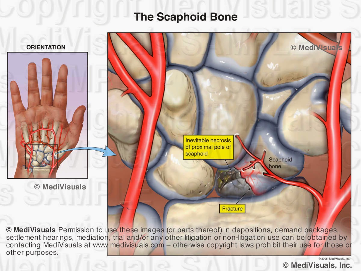
Like all organ systems, bones require a blood supply. If the blood supply is disrupted, the bone can die and collapse. This condition is known as avascular necrosis. The scaphoid is a bone in the wrist that is frequently affected by avascular necrosis. In the illustration below, a fracture is seen through the scaphoid and its blood supply.  As a result, the small fractured fragment loses its blood supply and over time, becomes necrotic. Treatment would likely involve fusion of the wrist bones. Other bones that are particularly vulnerable to avascular necrosis include the femoral head (hip joint) and the talus (ankle joint).


Ossification of the surrounding soft tissues is another complication of fractures. This condition is referred to as “heterotopic ossification” and/or “myositis ossificans”. With this condition, the bone cells that form to heal the fracture extend out into the surrounding tissues (see image below) resulting in pain and decreased range of motion. Treatment involves surgery to excise the excessive bone growth.


Wednesday, March 5, 2014

Understanding Traumatic Brain Injuries: "Mild" (less severe) to Severe - Part 2

 By: Robert Shepherd MS, Certified Medical Illustrator, President & CEO, MediVisuals Inc.

This article is a continuation of a two-part article on traumatic brain injury. Part 1 covered severe traumatic brain injury while part 2 addresses “mild” less severe traumatic brain injury. A person suffers a brain injury once every few seconds in the United States, with many going undiagnosed. Significant facts associated with these injuries include: 1) MRI or CT imaging studies not showing injuries, 2) the injured person might not think anything is wrong with them, and 3) physicians and others who did not know the patient prior to the traumatic brain injury may not appreciate the cognitive deficits and diagnose the condition. (Often, only persons who knew the injured person before the accident notice differences in personality, behavior, or cognitive function.)


During trauma, illustrated above, the brain impacts against the inside of the skull. Shearing injuries often occur because the gray and white matter are of different densities; therefore, the axons tear at the junction of the white and gray matter. The injuries can consist of torn or twisted axons, or the axons can pull away from their synapse.

Axonal injury can also occur without the head striking an object. This often occurs in collisions. During a sudden deceleration injury, the brain impacts the inside of the skull in a coup - contrecoup fashion, which means that the brain first impacts the area of the skull receiving the trauma and then impacts the area of the skull directly opposite of the trauma, as seen in the animation below. As a result, shock waves of the forces travel through the brain.


During sudden deceleration, the brain impacts on the hard jagged ridges of the base of the skull causing shearing forces, as depicted in the illustration below.


Blood vessels may also become torn or broken during a TBI, resulting in bleeding (see image below). An MRI or CT is not capable of detecting individual or even relatively large areas of axonal injury. Lesions detected by MRI or CT are typically areas of hemorrhage, if the hemorrhages are large enough.


Axons range in diameter from 1/4 of a micron to 10 microns while blood vessels range in diameter from 30 to 240 microns. If forces are sufficient to tear the much larger and resilient blood vessels (see illustration below), it is certain that numerous axons in the adjacent and other areas are torn as well. However, axons may be torn without injury and significant hemorrhage from nearby blood vessel is not torn, so the absence of findings on MRI or CT DO NOT RULE OUT traumatic brain injuries.


When hemorrhaging is not involved, traditional imaging studies, such as MRI or CT, are able to detect only large areas of axonal injury where thousands of axonal injuries create an area of abnormality large enough to be detected.

The loss of the sense of smell is an indicator of traumatic brain injury. The image below depicts the normal olfactory anatomy with the olfactory nerves extending through the cribiform plate and innervating the nasal passages. During trauma to the head, the forces can be great enough to sever the relatively large olfactory
nerves, which affects the sense of smell. Forces sufficient to injure the olfactory nerves are certainly sufficient to result in diffuse axonal injuries throughout the brain whether evident on imaging studies or not.


Problems with many functions (such as hearing, speech, and balance) following head trauma can result from injury to axons anywhere along the pathway involved in performing those function. For example, the ability to repeat a spoken word requires the proper function of the neural pathways for hearing and speaking.


Keys to detecting and proving “mild” less severe traumatic brain injuries are as follows:

1) Rely on changes of behavior and cognitive function as reported by family members, coworkers and friends. Casual examinations by a physician may not result in a diagnosis.

2) The absence of physical brain injuries on traditional MRI or CT DOES NOT RULE OUT brain injuries.

3) Correlation of traumatic forces with injury to the specific areas of the brain that control those functions is very important when proving a “mild” less severe traumatic brain injury.

Wednesday, February 26, 2014

Understanding Traumatic Brain Injuries: Mild (less severe) to Severe - Part 1

 By: Robert Shepherd MS, Certified Medical Illustrator, President & CEO, MediVisuals Inc.

Brain injuries are classified into two basic categories; those that are associated with obvious, incontestable evidence of intracranial injury and those that are not.

Obvious intracranial injuries include those with evidence of pathology within the brain itself intraparenchymal injuries as well as areas of bleeding around the brain but within the skull. The light area in the scan below indicates blood within the brain tissue, and the surrounding dark area shows associated edema. Both of these are considered to be intraparenchymal injuries. Contusions and hematomas found outside the skull are not considered intracranial but are frequently illustrated to help emphasize the force and direction of the trauma to the head.


The areas of hemorrhage shown in the illustration below are scattered around the junction between the grey and white matter of the brain, which is consistent with injuries from shear forces. The grey and white matter are of different densities, and when the brain impacts the skull during a traumatic event, the subsequent unequal movement between the two causes damage at their junction.

Head trauma can cause tearing of the blood vessels around the brain, which can result in areas of bleeding (hematomas). As the bleeding continues, the hematoma may expand to compress the brain tissue (as shown in the following illustration) and may require an emergency decompression. Significant compression must be relieved quickly in order to avoid further neurological damage and/or death.


Introducing a ventriculostomy catheter is one approach used to alleviate increased intracranial pressure. A hole is drilled directly into the patient's skull, and a catheter is advanced through the brain tissue into one of the ventricles. The catheter allows some of the cerebrospinal fluid to escape, thereby relieving the pressure on the brain itself.


In other situations, a craniotomy may be performed to allow the blood to be suctioned from the area surrounding the brain. A bone flap is created by first drilling three holes into the skull and then making cuts between them with a saw. This flap is removed and the blood is cleared from the areas above and beneath the dura. In some cases, the bone flap is not replaced if the brain is swollen or if there is a significant concern over recurrence of the hematoma. For a more detailed look at the craniotomy procedure, please review MediVisuals Craniotomy Surgery Animation.


© MediVisuals, Inc. - Permission to use any image (or parts thereof) posted on this blog in deposition, demand packages, settlement, hearings, mediation, trial, and/or any other litigation or non-litigation use can be obtained by contacting MediVisuals at www.medivisuals.com - otherwise copyright laws prohibit their use for those or other purposes.