MediVisuals

Scroll down to view blog archive

Scroll down to view blog archive
Showing posts with label ankle. Show all posts

Wednesday, May 14, 2014

Breaking Down Traumatic Arthritis

 By: Robert Shepherd MS, Certified Medical Illustrator, President & CEO, MediVisuals Inc.

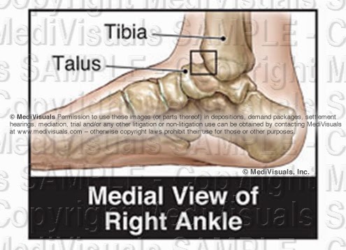
Fractures can result in several long term or permanent complications that can necessitate additional surgical procedures. One of the most common long term debilitating complications is traumatic arthritis.Traumatic arthritis can affect almost any moveable joint in the body. To explain traumatic arthritis more in depth, we will be focusing on the tibiotalar (ankle) joint, as shown in the illustration below.


 Buy on PI Exhibits NOW

As like most moveable joints, the tibiotalar joint consists of smooth articular bone covered by thick, shock-absorbing articular cartilage. 


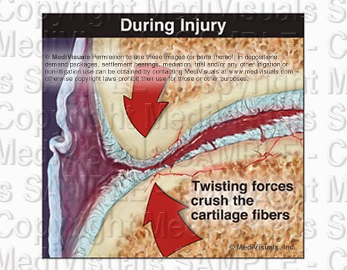
During trauma, the joint surfaces can be driven together resulting in injuries to the cartilage and microfractures of the articular surface (even without obvious intrarticular fracture). 


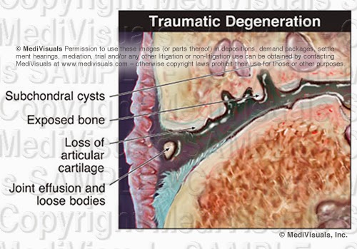
The bone and cartilage then undergo changes that result in the progressive breakdown of the joint. As the process advances, the joint becomes painful. In most cases, the only treatment options are joint replacement or fusion.


The acromioclavicular (AC) joint also frequently falls victim to traumatic arthritis. As the AC joint enlarges (hypertrophy) it impinges on the rotator cuff, which is referred to as subacromial impingement. This can cause irritation or tearing of the rotator cuff and is most often treated by AC joint resection and subacromial decompression.


Wednesday, April 9, 2014

Complications Associated with Fracture Injuries

 By: Robert Shepherd MS, Certified Medical Illustrator, President & CEO, MediVisuals Inc.

Several long term or permanent complications may result from a fracture injury. Traumatic arthritis may be one of the most common long term debilitating complications. (See the previously posted article "Breaking Down Traumatic Arthritis" for more information.) These complications may even require additional surgical procedures.

Normally, fractures begin healing by forming a callus which is then replaced by bone. With a nonunion (see image below), the callus is replaced by fibrous tissue instead of by bone. This is a painful condition that is most frequently treated by surgery to remove the fibrous tissue, debride the surrounding bone and re-approximate the remaining fragments (sometimes with bone graft material and fixation devices).


Malunion is another frequently occurring complication of fractures. With malunion, the bones heal, but they do so at an unusual angle that can effect range of motion and/or cause pain (see image below). Treatment typically involves refracturing or cutting the bones, realignment, and fixating them with hardware so that they heal in the correct position.


Intra-articular “step-offs” are a complication of intra-articular fractures. A “step-off” is a type of malunion in which the bone heals, but it heals so that the joint surfaces are not aligned in a smooth continuous surface. The illustration below shows a fracture through the patella that healed with a painful “step-off.”


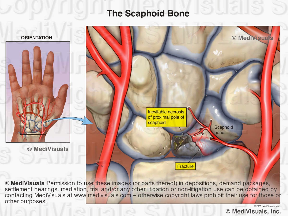
Like all organ systems, bones require a blood supply. If the blood supply is disrupted, the bone can die and collapse. This condition is known as avascular necrosis. The scaphoid is a bone in the wrist that is frequently affected by avascular necrosis. In the illustration below, a fracture is seen through the scaphoid and its blood supply.  As a result, the small fractured fragment loses its blood supply and over time, becomes necrotic. Treatment would likely involve fusion of the wrist bones. Other bones that are particularly vulnerable to avascular necrosis include the femoral head (hip joint) and the talus (ankle joint).


Ossification of the surrounding soft tissues is another complication of fractures. This condition is referred to as “heterotopic ossification” and/or “myositis ossificans”. With this condition, the bone cells that form to heal the fracture extend out into the surrounding tissues (see image below) resulting in pain and decreased range of motion. Treatment involves surgery to excise the excessive bone growth.


Wednesday, May 22, 2013

Intra-operative Trauma: The Overlooked Injuries

 By: Robert Shepherd MS, Certified Medical Illustrator, President & CEO, MediVisuals Inc.

The surgical trauma that a plaintiff has to undergo after the initial bodily injuries following a traumatic event are always major points of emphasis when arguing damages in a personal injury case.  This is certainly the situation with cases that involve broken bones that require invasive surgical procedures to realign broken bone fragments ("reduce") and secure ("fixate") the bones with hardware to keep them properly aligned during healing.  Too often, however, the emphasis is solely on the effects on the bones from these "Open Reduction and Internal Fixation" (ORIF) procedures, and very little emphasis is placed on the surgical disruption of the soft tissues that takes place during these procedures.

In a case involving ORIF of a distal fibula (a.k.a. lateral malleolus) fracture, in order to emphasize the surgical trauma endured by a plaintiff, an attorney may have a visual prepared of a postoperative X-ray.  The visual may consist of only a postoperative X-ray or a print of the X-ray with a corresponding illustration (see the below figure). 

The above images are certainly helpful, but fail to address the intra-operative trauma to the soft tissues that is required to gain access to the bone fragments.  For that purpose, intra-operative illustrations that truthfully depict the soft tissue disruption should be considered (see the below figure) or even an animation showing the procedures such as the one at this link: http://www.medivisuals.com/fibularplatingORIF.aspx
Illustrations or animations that at least touch on the soft tissue disruption allow testifying physicians the opportunity to explain the many tissues traumatized during the procedure and allow insurance adjustors, mediators, and jurors an opportunity to take these additional injuries into consideration when determining the severity of a plaintiff's entire injuries.


Many attorneys considering realistic illustrations such as the one above, express a concern that judges may not allow the images to be used because they are too "graphic" or "inflammatory".  Certainly, counsel should make themselves aware and consider the preferences of certain jurisdictions and specific judges before determining whether an illustration should be developed that realistically depicts injuries or whether diagrammatic (cartoon-like) illustrations should be developed instead.  There are a number of very good arguments to support the use of "realistic" illustrations over "cartoons".  Those arguments as well as other discussions regarding illustration styles will be addressed in future blogs.
© 2012 MediVisuals, Inc. 

Thursday, July 5, 2012

Breaking Down Traumatic Arthritis

 By: Robert Shepherd MS, Certified Medical Illustrator, President & CEO, MediVisuals Inc.

Fractures can result in several long term or permanent complications that can necessitate additional surgical procedures. One of the most common long term debilitating complications is traumatic arthritis.Traumatic arthritis can affect almost any moveable joint in the body. To explain traumatic arthritis more in depth, we will be focusing on the tibiotalar (ankle) joint, as shown in the illustration below.
As like most moveable joints, the tibiotalar joint consists of smooth articular bone covered by thick, shock-absorbing articular cartilage.
During trauma, the joint surfaces can be driven together resulting in injuries to the cartilage and microfractures of the articular surface (even without obvious intrarticular fracture).
The bone and cartilage then undergo changes that result in the progressive breakdown of the joint. As the process advances, the joint becomes painful. In most cases, the only treatment options are joint replacement or fusion.
The acromioclavicular (AC) joint also frequently falls victim to traumatic arthritis. As the AC joint enlarges (hypertrophy) it impinges on the rotator cuff, which is referred to as subacromial impingement. This can cause irritation or tearing of the rotator cuff and is most often treated byAC joint resection and subacromial decompression.