MediVisuals

Scroll down to view blog archive

Scroll down to view blog archive
Showing posts with label medical exhibit. Show all posts

Wednesday, April 9, 2014

Complications Associated with Fracture Injuries

 By: Robert Shepherd MS, Certified Medical Illustrator, President & CEO, MediVisuals Inc.

Several long term or permanent complications may result from a fracture injury. Traumatic arthritis may be one of the most common long term debilitating complications. (See the previously posted article "Breaking Down Traumatic Arthritis" for more information.) These complications may even require additional surgical procedures.

Normally, fractures begin healing by forming a callus which is then replaced by bone. With a nonunion (see image below), the callus is replaced by fibrous tissue instead of by bone. This is a painful condition that is most frequently treated by surgery to remove the fibrous tissue, debride the surrounding bone and re-approximate the remaining fragments (sometimes with bone graft material and fixation devices).


Malunion is another frequently occurring complication of fractures. With malunion, the bones heal, but they do so at an unusual angle that can effect range of motion and/or cause pain (see image below). Treatment typically involves refracturing or cutting the bones, realignment, and fixating them with hardware so that they heal in the correct position.


Intra-articular “step-offs” are a complication of intra-articular fractures. A “step-off” is a type of malunion in which the bone heals, but it heals so that the joint surfaces are not aligned in a smooth continuous surface. The illustration below shows a fracture through the patella that healed with a painful “step-off.”


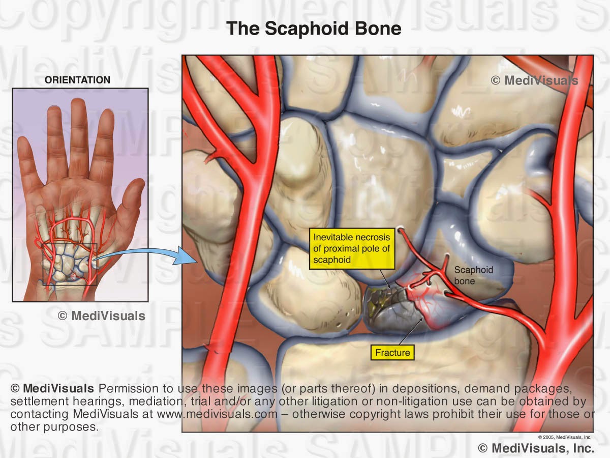
Like all organ systems, bones require a blood supply. If the blood supply is disrupted, the bone can die and collapse. This condition is known as avascular necrosis. The scaphoid is a bone in the wrist that is frequently affected by avascular necrosis. In the illustration below, a fracture is seen through the scaphoid and its blood supply.  As a result, the small fractured fragment loses its blood supply and over time, becomes necrotic. Treatment would likely involve fusion of the wrist bones. Other bones that are particularly vulnerable to avascular necrosis include the femoral head (hip joint) and the talus (ankle joint).


Ossification of the surrounding soft tissues is another complication of fractures. This condition is referred to as “heterotopic ossification” and/or “myositis ossificans”. With this condition, the bone cells that form to heal the fracture extend out into the surrounding tissues (see image below) resulting in pain and decreased range of motion. Treatment involves surgery to excise the excessive bone growth.


Wednesday, February 26, 2014

Understanding Traumatic Brain Injuries: Mild (less severe) to Severe - Part 1

 By: Robert Shepherd MS, Certified Medical Illustrator, President & CEO, MediVisuals Inc.

Brain injuries are classified into two basic categories; those that are associated with obvious, incontestable evidence of intracranial injury and those that are not.

Obvious intracranial injuries include those with evidence of pathology within the brain itself intraparenchymal injuries as well as areas of bleeding around the brain but within the skull. The light area in the scan below indicates blood within the brain tissue, and the surrounding dark area shows associated edema. Both of these are considered to be intraparenchymal injuries. Contusions and hematomas found outside the skull are not considered intracranial but are frequently illustrated to help emphasize the force and direction of the trauma to the head.


The areas of hemorrhage shown in the illustration below are scattered around the junction between the grey and white matter of the brain, which is consistent with injuries from shear forces. The grey and white matter are of different densities, and when the brain impacts the skull during a traumatic event, the subsequent unequal movement between the two causes damage at their junction.

Head trauma can cause tearing of the blood vessels around the brain, which can result in areas of bleeding (hematomas). As the bleeding continues, the hematoma may expand to compress the brain tissue (as shown in the following illustration) and may require an emergency decompression. Significant compression must be relieved quickly in order to avoid further neurological damage and/or death.


Introducing a ventriculostomy catheter is one approach used to alleviate increased intracranial pressure. A hole is drilled directly into the patient's skull, and a catheter is advanced through the brain tissue into one of the ventricles. The catheter allows some of the cerebrospinal fluid to escape, thereby relieving the pressure on the brain itself.


In other situations, a craniotomy may be performed to allow the blood to be suctioned from the area surrounding the brain. A bone flap is created by first drilling three holes into the skull and then making cuts between them with a saw. This flap is removed and the blood is cleared from the areas above and beneath the dura. In some cases, the bone flap is not replaced if the brain is swollen or if there is a significant concern over recurrence of the hematoma. For a more detailed look at the craniotomy procedure, please review MediVisuals Craniotomy Surgery Animation.


© MediVisuals, Inc. - Permission to use any image (or parts thereof) posted on this blog in deposition, demand packages, settlement, hearings, mediation, trial, and/or any other litigation or non-litigation use can be obtained by contacting MediVisuals at www.medivisuals.com - otherwise copyright laws prohibit their use for those or other purposes. 


Wednesday, May 22, 2013

Intra-operative Trauma: The Overlooked Injuries

 By: Robert Shepherd MS, Certified Medical Illustrator, President & CEO, MediVisuals Inc.

The surgical trauma that a plaintiff has to undergo after the initial bodily injuries following a traumatic event are always major points of emphasis when arguing damages in a personal injury case.  This is certainly the situation with cases that involve broken bones that require invasive surgical procedures to realign broken bone fragments ("reduce") and secure ("fixate") the bones with hardware to keep them properly aligned during healing.  Too often, however, the emphasis is solely on the effects on the bones from these "Open Reduction and Internal Fixation" (ORIF) procedures, and very little emphasis is placed on the surgical disruption of the soft tissues that takes place during these procedures.

In a case involving ORIF of a distal fibula (a.k.a. lateral malleolus) fracture, in order to emphasize the surgical trauma endured by a plaintiff, an attorney may have a visual prepared of a postoperative X-ray.  The visual may consist of only a postoperative X-ray or a print of the X-ray with a corresponding illustration (see the below figure). 

The above images are certainly helpful, but fail to address the intra-operative trauma to the soft tissues that is required to gain access to the bone fragments.  For that purpose, intra-operative illustrations that truthfully depict the soft tissue disruption should be considered (see the below figure) or even an animation showing the procedures such as the one at this link: http://www.medivisuals.com/fibularplatingORIF.aspx
Illustrations or animations that at least touch on the soft tissue disruption allow testifying physicians the opportunity to explain the many tissues traumatized during the procedure and allow insurance adjustors, mediators, and jurors an opportunity to take these additional injuries into consideration when determining the severity of a plaintiff's entire injuries.


Many attorneys considering realistic illustrations such as the one above, express a concern that judges may not allow the images to be used because they are too "graphic" or "inflammatory".  Certainly, counsel should make themselves aware and consider the preferences of certain jurisdictions and specific judges before determining whether an illustration should be developed that realistically depicts injuries or whether diagrammatic (cartoon-like) illustrations should be developed instead.  There are a number of very good arguments to support the use of "realistic" illustrations over "cartoons".  Those arguments as well as other discussions regarding illustration styles will be addressed in future blogs.

© 2012 MediVisuals, Inc. 

Disc-Osteophyte Complex Explained

 By: Robert Shepherd MS, Certified Medical Illustrator, President & CEO, MediVisuals Inc.

Individuals who develop new or suddenly worsening symptoms consistent with nerve root or spinal cord impingement following a traumatic event are sometimes diagnosed with “disc-osteophyte complexes”. The term “disc-osteophyte complex” generally refers to abnormal extension of intervertebral disc material that accompanies immediately adjacent osteophyte formation at the vertebral body margin (see the below figure). It is important to note (as shown in the illustrations) that the disc almost always extends further than the osteophytes into the neural foramen or spinal canal to irritate or impinge upon nerve roots or the spinal cord.


Occasionally, individuals who are evaluated shortly after a traumatic event are found to have disc-osteophyte complexes. Because a minimum of several weeks is required for osteophytes to form as a result of a traumatic event, defendant insurance companies may argue that the presence of osteophytes so soon after the traumatic event in question may prove that the plaintiff’s injuries preexisted the traumatic event. Since it is the disc pathology extending beyond the osteophytes that is the actual cause of the nerve root or spinal cord irritation and inflammation, the defense’s arguments are not valid. As shown in the illustrations below, the sequence of events that typically takes place in these cases is that the plaintiff had minimally symptomatic or asymptomatic disc osteophytes prior to the traumatic event in question. During the traumatic event, the disc sustains trauma that results in worsening of the disc pathology while the osteophyte portion of the osteophyte/disc complex remains essentially unchanged. This worsening of the disc pathology in turn results in new or increased irritation or impingement of the neural elements.


© 2012 MediVisuals, Inc


Thursday, June 14, 2012

Medical Legal Illustration and Animation: Cross Sections

 By: Robert Shepherd MS, Certified Medical Illustrator, President & CEO, MediVisuals Inc.

Long before science had advanced to allow imaging of the body in sectional views by computed tomography (CT) and magnetic resonance imaging (MRI), medical illustrators were illustrating the body in sectional views because these views are the best way to appreciate some anatomical relationships. 

Medical illustrators, physicians, and others who have studied anatomy are familiar with sectional views of the body and appreciate the value of these views in explaining the relationship of anatomical structures.  However, accomplished and well respected jury consultants and non-medical illustrator legal graphics experts have expressed concerns that sectional views may be difficult for some jury members to understand.  These individuals' opinions are valuable to those of us in the legal graphics business, and I agree with their opinions that, when other views can communicate a particular relationship message equally as well or better, sectional views should be avoided. I also believe most of these experts will agree that there are times and places in which sectional views of anatomy are the best way to appreciate some anatomical relationships.  Granted, there have been times when we have been working on specific cases and experts have insisted that sectional views be absolutely and unconditionally avoided.  Unfortunately, in these situations the experts were unable to suggest a more effective view to communicate the relevant anatomical relationships (at least in a way that was practical in terms of time and expense). That being the case, sometimes the sectional views were used despite the input of the experts, and at other times, the relationships of the structures had to be explained without the benefits of graphics.
A way to perhaps explain how sectional views help decision makers appreciate relevant anatomical and pathological relationships is to compare them to aerial views or photographs of the scene of a collision.  Space is defined in three planes.  Only two of these planes can effectively be demonstrated in a two-dimensional rendering.  For example, aerial views have long been used to help explain the positions of vehicles and structures that simply can't be appreciate from "street views".   When viewing the scene of a collision from a "street view", one can appreciate vertical and horizontal distances, but not depth; distances close to and far from the viewer's perspective are very difficult to appreciate (see the below figures).  By comparison, when viewing an operative site through a "surgeon's view", vertical and horizontal distances can be appreciated, but the depth of the incision and the relationships of the various structures within and around the incision are very difficult or impossible to appreciate.


The "aerial view" of the collision scene allows the viewer to appreciate distances in two geographical planes as well (distances right and left, and toward and away from the "street view," but the ability to appreciate up and down is lost).  Also, the locations of relevant structures or vehicles that may have been obstructed by nearby structures (such as buildings trees, signs, or other vehicles) can now be appreciated.  Similarly, a sectional view of anatomy can help decision makers appreciate depth relationships of structures.  Or, a sectional view of a step in a surgical illustration can allow the viewer to appreciate the depth of the surgery as well as the additional structures that may have been injured (or at risk of injury) during the invasive procedure.  These specific depth  relationships could not be appreciated from the "surgeon's view" of the same surgery shown in the above illustration.


Exhibits developed to help explain the invasive nature of a surgery and the disruption of the soft tissues during operative procedures are critical.  For that reason, sectional views are critical in aiding a testifying physician to explain these issues.  For example, the exhibit panel that demonstrates an anterior cervical discectomy and fusion (ACDF) that does not include a cross-section through the neck fails to emphasize the depth of the incision and disruption of tissues (essentially all the way to the center of the neck).  This depth simply cannot be appreciated in a "surgeon's view".
In order to appreciate cross-sections, orientation views that show the level and direction of the section are helpful (see below), or when time, budget, and presentation format (digital as opposed to a physical panel) allow, a short animation showing the sectional view actually coming out of the orientation view such as MediVisuals' "Scan SelectorTM" can be used.