MediVisuals

Scroll down to view blog archive

Scroll down to view blog archive
Showing posts with label intervertebral disc. Show all posts

Tuesday, November 12, 2013

Understanding the Osteophyte/Disc Complex in Spinal Trauma

By: Robert Shepherd MS, Certified Medical Illustrator, President & CEO, MediVisuals Inc.

A traumatic event causing injury to an intervertebral disc may also cause subtle injuries to the bones around the disc. During an extreme lateral flexion injury (shown in the image below), the edges of the bone are driven together, injuring both the disc and the bone. As the bone/disc junction heals, overgrowths referred to as osteophytes may form.


Osteophytes take weeks or months to develop following a traumatic event; therefore, any osteophytes that are present soon after a traumatic event are likely pre-existing.


The osteophytes themselves may compress the neural elements as in the illustration above; however, in most situations, the osteophytes are a part of an OSTEOPHYTE/DISC COMPLEX. This is when the osteophytes and disc extend beyond their normal limits and compress the neural elements (spinal cord, nerve roots). In cases where osteophytes may have pre-existed a traumatic event, worsening of the disc bulge could occur following the trauma, resulting in new or aggravated symptoms.

Sometimes disc and ligament injuries occur on the same side as the force of impact. Other times, they occur on the opposite side. Injuries to the disc on the same side as the force are the result of stretching and tearing forces. On the opposite side, compression forces result in tears and micro fractures of the tissues and bones. (see illustration below). Osteophytes and facet hypertrophy can also form following injuries to intervertebral discs and ligaments. Injuries to the discs and ligaments result in instability and excessive motion of the joints that, in turn, results in constant trauma to the bone/disc and ligament junctions. This ongoing trauma results in overgrowth of the bones as it continually cycles through episodes of healing and reinjury.

Wednesday, May 22, 2013

How a Disc "Bulge" is Different From a "Herniation" (Intervertebral Disc Pathology, Part 1 of 3)

 By: Robert Shepherd MS, Certified Medical Illustrator, President & CEO, MediVisuals Inc.


It is difficult to appreciate the subtle differences between the various types or severities of intervertebral disc injuries that result in them being defined as bulges, herniations, protrusions, extrusions, etc.  The way disc pathology is defined may even vary from physician to physician—perhaps primarily due to the fact that, prior to 1995, many physicians’ professional societies used different criteria to define the various classifications of disc injuries.  In 1995, a joint undertaking by representatives from the North American Spine Society, the American Society of Spine Radiology, and the American Society of Neuroradiology worked together to develop a more widely accepted and used system to define disc pathology as published in "Nomenclature and Classification of Lumbar Disc Pathology”.

This will be the first of three blogs dedicated to helping explain the definitions of disc pathology as recommended by the 1995 combined task force. This blog will focus on the difference between "bulges" and "herniations". Topics to be discussed in future articles are differences between a "Herniated Disc" and an "Annular Tear" and the difference between "Protrusions" and  "Extrusions".

In the image below, a normal disc is shown in comparison to the two types of intervertebral disc injuries covered in this article: "Bulges" and "Herniations". Disc "Bulges", in general, are defined by the presence of disc material beyond the normal margins around at least 50% of the disc's circumference. A "Herniation" is defined as displacement of disc material beyond the limits of the intervertebral disc space that extends less than 50% around the circumference of the disc. The displacement material can consist of the nucleus, the annulus, or parts of both. This is significant in personal injury litigation because the defense often places a great deal of emphasis on whether disc pathology is defined as a "bulge" or "herniation" when determining the severity of an injury. However, a "bulge" can actually impinge nerve roots or the spinal cord to a more severe degree than a "herniation".


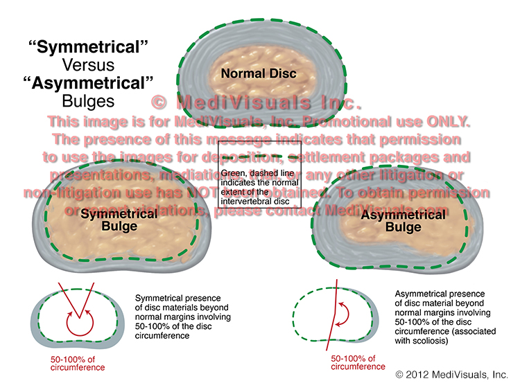
The next image compares the normal disc to two different types of disc "Bulges". A "Bulge" is defined as "Symmetrical" when the right and left sides of the herniation more or less mirror each other.  A bulge is "Asymmetrical" when the bulge is more severe on one side when compared to the other.


Finally, the below image shows a normal disc as compared to two types of "Herniations". A "Broad-Based" herniation is defined as disc material extending beyond its normal limits in an area between 25 and 50% of the disc's circumference.  A "Focal" herniation is one involving extension of disc material beyond its normal limits in less than 25% of its circumference.


Annular Tears and Fissures (Intervertebral Disc Pathology, Part 2 of 3)

 By: Robert Shepherd MS, Certified Medical Illustrator, President & CEO, MediVisuals Inc.

This blog is a follow-up referencing language and labels used by health professionals to describe various types of intervertebral disc pathology as defined by a 1995 joint undertaking by representatives from the North American Spine Society, American Society of Spine Radiology and American Society of Neuroradiology.  As a result of their efforts, a more uniform and widely accepted use of nomenclature to define intervertebral disc pathology was developed and published in "Nomenclature and Classification of Lumbar Disc Pathology".

A previous blog dealt with "Symmetrical" and "Asymmetrical" disc "bulge" and "Broad-based" v. "Focal" Herniations.  The first disc pathology term discussed in this blog is "Annular Tear".  This is essentially synonymous with "Annular Fissure," with perhaps "Fissure" being preferable over "Tear" because "Tear" may imply that the pathology was the result of some sort of traumatic event, and this specific pathology can occur without necessarily being the result of trauma.  Annular Tears/Fissures, as seen in the below figure, can occur without fitting the definition of a "Herniation" (disc material extruding beyond its normal boundaries).  As seen in the below illustrations, the fibers of the annulus can be torn with nucleus protruding into the annulus but without the annulus or nucleus extending beyond the bordering vertebral bodies.  By contrast, when "Anular Tears/Fissures" result in disc material extending beyond its normal boundaries, the disc pathology is typically referred to simply as a "Herniation" without a reference to the presence of an Annular Tear/Fissure."


Another blog will be coming soon regarding disc "Protrusions" and "Extrusions".

© 2012 MediVisuals, Inc.

Protrusions Versus Extrusions (Intervertebral Disc Pathology, Part 3 of 3)

 By: Robert Shepherd MS, Certified Medical Illustrator, President & CEO, MediVisuals Inc.


This blog is the third in a series referencing language and labels used by health professionals to describe intervertebral disc pathology as defined by a 1995 joint undertaking by representatives from the North American Spine Society, the American Society of Spine Radiology and the American Society of Neuroradiology.  As a result of their efforts, a more uniform and widely accepted use of nomenclature to define intervertebral disc pathology was developed and published in "Nomenclature and Classification of Lumbar Disc Pathology".

The first blog in the series dealt with "Bulges" v. "Herniations", "Symmetrical" and "Asymmetrical" disc bulges and "Broad-based" v. "Focal" herniations.  The second blog addressed "Anular Tears and Fissures".  This blog addresses the use of "Protrusion" and "Extrusion" to describe intervertebral disc herniations.

"Protrusion" and "Extrusion" are essentially used to further classify types of disc herniations. The term "Protrusion" refers to a disc herniation in which the portion of disc material that is outside the normal confines of the disc space is equal to or less than its aperture where the disc material extrudes from the parent disc.   Examples of disc "Protrusions" and "Extrusions" are shown in the below images. 

The image to the left shows a disc "Protrusion".  Note how the superior and inferior dimensions of the disc material that protrudes from its normal confines (highlighted by the arrow on the left) is not as great as the area where the protruding disc material actually exits its normal confines and boundaries (represented by the arrow on the right in the image).  By comparison, the two illustrations to the right show two different disc "Extrusions".  Note how the dimensions of the protruding disc material are greater than the point where it exits its normal confines. 

It is important to appreciate that disc "Protrusions" and "Extrusions" are terms that may be used to further describe "Broad-based" or "Focal" herniations.  For example, the disc pathology referred to in the above illustration as a "Protrusion" could also be "Broad-Based," if it extends between 25 and 50% of the distance around the circumference of the vertebral body.  Similarly, the disc pathology shown in the illustrations referred to as "Extrusions" could also be referred to as "Focal" if extending less than 25% of the distance around the circumference of the vertebral body (see blog from 08/24/11 for further clarification between "Broad-based" and "Focal" herniations).
© 2012 MediVisuals, Inc.

Disc-Osteophyte Complex Explained

 By: Robert Shepherd MS, Certified Medical Illustrator, President & CEO, MediVisuals Inc.

Individuals who develop new or suddenly worsening symptoms consistent with nerve root or spinal cord impingement following a traumatic event are sometimes diagnosed with “disc-osteophyte complexes”. The term “disc-osteophyte complex” generally refers to abnormal extension of intervertebral disc material that accompanies immediately adjacent osteophyte formation at the vertebral body margin (see the below figure). It is important to note (as shown in the illustrations) that the disc almost always extends further than the osteophytes into the neural foramen or spinal canal to irritate or impinge upon nerve roots or the spinal cord.


Occasionally, individuals who are evaluated shortly after a traumatic event are found to have disc-osteophyte complexes. Because a minimum of several weeks is required for osteophytes to form as a result of a traumatic event, defendant insurance companies may argue that the presence of osteophytes so soon after the traumatic event in question may prove that the plaintiff’s injuries preexisted the traumatic event. Since it is the disc pathology extending beyond the osteophytes that is the actual cause of the nerve root or spinal cord irritation and inflammation, the defense’s arguments are not valid. As shown in the illustrations below, the sequence of events that typically takes place in these cases is that the plaintiff had minimally symptomatic or asymptomatic disc osteophytes prior to the traumatic event in question. During the traumatic event, the disc sustains trauma that results in worsening of the disc pathology while the osteophyte portion of the osteophyte/disc complex remains essentially unchanged. This worsening of the disc pathology in turn results in new or increased irritation or impingement of the neural elements.


© 2012 MediVisuals, Inc