MediVisuals

Scroll down to view blog archive

Scroll down to view blog archive
Showing posts with label discogenic pain. Show all posts

Wednesday, August 6, 2014

Discogenic Pain - Back Pain and Radiculopathy Without Evidence of Significant Disc Injury

 By: Robert Shepherd MS, Certified Medical Illustrator, President & CEO, MediVisuals Inc.

In determining if a person's pain may be related to some sort of intervertebral disc pathology, a great deal of emphasis is placed upon imaging studies showing evidence of mechanical compression of a nerve root by abnormal posterior displacement of a disc (i.e. bulge, protrusion, herniation, etc.) as portrayed in the below illustration.


In cases where clear mechanical compression of the nerve roots is not shown in imaging studies, some are quick to argue that any pain emanating from the area is either exaggerated or entirely contrived. However, a person can experience pain consistent with mechanical compression of a nerve root without having any significant disc pathology. This is because the spine is encircled with a meshwork of nerves that are much too small to be seen on CT or MRI (see the below figure). The sinuvertebral nerves surround and penetrate the intervertebral discs.
When injuries to a disc are more subtle, the sinuvertebral nerves may detect the injuries and send pain signals to the brain where they are interpreted as pain (see figure). The pain may be limited to the area of the back, or a pain perception phenomenon know as "pain referral" (confusion of the origin of pain signals by the brain) may result in the person experiencing very real pain consistent with radicular pain from mechanical nerve root compression by a severely herniated disk.
Another common cause of pain consistent with nerve root mechanical compression is chemical irritation or inflammation of the nerve root. Chemical irritation of a nerve root often results from the release of chemicals following a more subtle disc injury (see the below figure). These chemicals irritate and inflame the nerve root and surrounding tissues, resulting in the perception of pain consistent with an injury to the disc and mechanical compression of the nerve root. Even after resolution of chemical irritation and inflammation, scar tissue may develop that binds the nerve root (often undetectable on CT or MRI). This scarring can cause permanent debilitating pain that may require surgical intervention.

© MediVisuals, Inc. - Permission to use any image (or parts thereof) posted on this blog in depositions, demand packages, settlement hearings, mediation, trial, and/or any other litigation or non-litigation use can be obtained by contacting MediVisuals at trose@medivisuals.com – otherwise copyright laws prohibit their use for those or other purposes. 

Wednesday, October 23, 2013

Discogenic Pain - My Client Has Pain but No Disc Herniation

 By: Robert Shepherd MS, Certified Medical Illustrator, President & CEO, MediVisuals Inc.

Defense counsel, in personal injury cases involving spinal disc injuries, place a great deal of importance on the large neural structures, such as the nerve roots and spinal cord, but often times fail to appreciate the significance of the many smaller nerves around the spine. As the illustration below demonstrates, the spinal canal and discs are covered with a meshwork of nerves.  In some people, these nerves can be far more sensitive than they are in others.


Direct compression of an exiting nerve root (see illustration below) is widely appreciated to cause local, as well as, radicular pain and weakness.


However, if a physician's interpretation of a plaintiff's radiology films is that the films show only a bulge that does not compress the nerve root, the problem then becomes to prove that the plaintiff’s pain and weakness are not simply "fabricated".

Discogenic pain is a very likely explanation for local and radicular back pain. The disc itself has numerous sensory nerves called the sinuvertebral nerves. (see illustration below) With an injury to and/or a breakdown of the disc, these nerve endings are also damaged and send pain impulses through the spinal nerve roots.


Another explanation for pain and weakness without direct compression is attributable to chemical irritation of the nerve root due to the breakdown of the nearby disc. This occurs because, as the disc breaks down, chemicals and inflammation irritate the nearby nerve root causing pain and weakness, just as if the nerve root were compressed. (see illustration below)


In review, pain signals from the nerve root whether due to:
- Direct Compression
- Injury to the sinuvertebral nerves
- Chemical irritation of the nerve roots
 . . . and are carried to the brain and interpreted in the same way.

Wednesday, May 22, 2013

How a Disc "Bulge" is Different From a "Herniation" (Intervertebral Disc Pathology, Part 1 of 3)

 By: Robert Shepherd MS, Certified Medical Illustrator, President & CEO, MediVisuals Inc.


It is difficult to appreciate the subtle differences between the various types or severities of intervertebral disc injuries that result in them being defined as bulges, herniations, protrusions, extrusions, etc.  The way disc pathology is defined may even vary from physician to physician—perhaps primarily due to the fact that, prior to 1995, many physicians’ professional societies used different criteria to define the various classifications of disc injuries.  In 1995, a joint undertaking by representatives from the North American Spine Society, the American Society of Spine Radiology, and the American Society of Neuroradiology worked together to develop a more widely accepted and used system to define disc pathology as published in "Nomenclature and Classification of Lumbar Disc Pathology”.

This will be the first of three blogs dedicated to helping explain the definitions of disc pathology as recommended by the 1995 combined task force. This blog will focus on the difference between "bulges" and "herniations". Topics to be discussed in future articles are differences between a "Herniated Disc" and an "Annular Tear" and the difference between "Protrusions" and  "Extrusions".

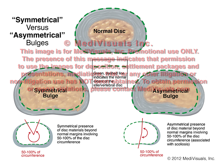
In the image below, a normal disc is shown in comparison to the two types of intervertebral disc injuries covered in this article: "Bulges" and "Herniations". Disc "Bulges", in general, are defined by the presence of disc material beyond the normal margins around at least 50% of the disc's circumference. A "Herniation" is defined as displacement of disc material beyond the limits of the intervertebral disc space that extends less than 50% around the circumference of the disc. The displacement material can consist of the nucleus, the annulus, or parts of both. This is significant in personal injury litigation because the defense often places a great deal of emphasis on whether disc pathology is defined as a "bulge" or "herniation" when determining the severity of an injury. However, a "bulge" can actually impinge nerve roots or the spinal cord to a more severe degree than a "herniation".


The next image compares the normal disc to two different types of disc "Bulges". A "Bulge" is defined as "Symmetrical" when the right and left sides of the herniation more or less mirror each other.  A bulge is "Asymmetrical" when the bulge is more severe on one side when compared to the other.


Finally, the below image shows a normal disc as compared to two types of "Herniations". A "Broad-Based" herniation is defined as disc material extending beyond its normal limits in an area between 25 and 50% of the disc's circumference.  A "Focal" herniation is one involving extension of disc material beyond its normal limits in less than 25% of its circumference.


Annular Tears and Fissures (Intervertebral Disc Pathology, Part 2 of 3)

 By: Robert Shepherd MS, Certified Medical Illustrator, President & CEO, MediVisuals Inc.

This blog is a follow-up referencing language and labels used by health professionals to describe various types of intervertebral disc pathology as defined by a 1995 joint undertaking by representatives from the North American Spine Society, American Society of Spine Radiology and American Society of Neuroradiology.  As a result of their efforts, a more uniform and widely accepted use of nomenclature to define intervertebral disc pathology was developed and published in "Nomenclature and Classification of Lumbar Disc Pathology".

A previous blog dealt with "Symmetrical" and "Asymmetrical" disc "bulge" and "Broad-based" v. "Focal" Herniations.  The first disc pathology term discussed in this blog is "Annular Tear".  This is essentially synonymous with "Annular Fissure," with perhaps "Fissure" being preferable over "Tear" because "Tear" may imply that the pathology was the result of some sort of traumatic event, and this specific pathology can occur without necessarily being the result of trauma.  Annular Tears/Fissures, as seen in the below figure, can occur without fitting the definition of a "Herniation" (disc material extruding beyond its normal boundaries).  As seen in the below illustrations, the fibers of the annulus can be torn with nucleus protruding into the annulus but without the annulus or nucleus extending beyond the bordering vertebral bodies.  By contrast, when "Anular Tears/Fissures" result in disc material extending beyond its normal boundaries, the disc pathology is typically referred to simply as a "Herniation" without a reference to the presence of an Annular Tear/Fissure."


Another blog will be coming soon regarding disc "Protrusions" and "Extrusions".

© 2012 MediVisuals, Inc.

Protrusions Versus Extrusions (Intervertebral Disc Pathology, Part 3 of 3)

 By: Robert Shepherd MS, Certified Medical Illustrator, President & CEO, MediVisuals Inc.


This blog is the third in a series referencing language and labels used by health professionals to describe intervertebral disc pathology as defined by a 1995 joint undertaking by representatives from the North American Spine Society, the American Society of Spine Radiology and the American Society of Neuroradiology.  As a result of their efforts, a more uniform and widely accepted use of nomenclature to define intervertebral disc pathology was developed and published in "Nomenclature and Classification of Lumbar Disc Pathology".

The first blog in the series dealt with "Bulges" v. "Herniations", "Symmetrical" and "Asymmetrical" disc bulges and "Broad-based" v. "Focal" herniations.  The second blog addressed "Anular Tears and Fissures".  This blog addresses the use of "Protrusion" and "Extrusion" to describe intervertebral disc herniations.

"Protrusion" and "Extrusion" are essentially used to further classify types of disc herniations. The term "Protrusion" refers to a disc herniation in which the portion of disc material that is outside the normal confines of the disc space is equal to or less than its aperture where the disc material extrudes from the parent disc.   Examples of disc "Protrusions" and "Extrusions" are shown in the below images. 

The image to the left shows a disc "Protrusion".  Note how the superior and inferior dimensions of the disc material that protrudes from its normal confines (highlighted by the arrow on the left) is not as great as the area where the protruding disc material actually exits its normal confines and boundaries (represented by the arrow on the right in the image).  By comparison, the two illustrations to the right show two different disc "Extrusions".  Note how the dimensions of the protruding disc material are greater than the point where it exits its normal confines. 

It is important to appreciate that disc "Protrusions" and "Extrusions" are terms that may be used to further describe "Broad-based" or "Focal" herniations.  For example, the disc pathology referred to in the above illustration as a "Protrusion" could also be "Broad-Based," if it extends between 25 and 50% of the distance around the circumference of the vertebral body.  Similarly, the disc pathology shown in the illustrations referred to as "Extrusions" could also be referred to as "Focal" if extending less than 25% of the distance around the circumference of the vertebral body (see blog from 08/24/11 for further clarification between "Broad-based" and "Focal" herniations).
© 2012 MediVisuals, Inc.