MediVisuals

Scroll down to view blog archive

Scroll down to view blog archive
Showing posts with label disc injury. Show all posts

Friday, May 16, 2014

Disc Herniation and Other Disc Injuries

By: Robert Shepherd MS, Certified Medical Illustrator, President & CEO, MediVisuals Inc.

The term “degenerated disc” is generally used to describe a disc in the early degenerative process. It is the beginning of a progressive break down of the disc. This condition can be initiated or accelerated by a traumatic event.


A disc bulge is a more advanced collapse of the disc to the point that the disc expands beyond its normal contour. It may or may not impinge on the neural structures within the spinal canal or neural foramina. Similarly, a disc bulge can be the immediate or delayed result of a traumatic event, or a traumatic event may exacerbate a preexisting, stable disc bulge.


A subligamentous herniation is one in which nucleus pulposus has extended through the annulus fibrosus, but has not gone through the posterior longitudinal ligament.


The term “herniation” is generally used when the nucleus has completely extruded through the annulus fibrosus and posterior longitudinal ligament

However, it does not matter what the disc pathology is labeled, if it impinges upon, or irritates the neural components, it is a significant injury that will likely require some type of invasive procedure to correct. 

The disc can either be injured by an immediate tear of the annulus fibrosus and extrusion of the nucleus pulposus during a traumatic event, or they can be the result of a much more gradual process. In order to understand the gradual breakdown of the disc, one must first understand a little of the physiology of a disc. The inner disc relies on exchange of fluid, nutrients and oxygen through the end plate of the adjacent vertebral bodies.

During a traumatic event, the endplate may become injured resulting in interference with that exchange.
As a result of the inability of the disc to obtain the fluid, nutrients, and oxygen it needs, the disc gradually begins to break down - becoming a degenerated disc, followed by a bulging disc, and eventually to a herniated disc. The amount of time involved with the process of the disc breakdown is related to the severity of the initial disc injury.


Therefore, if a disc injury is not evident until weeks or months after a traumatic event, it does not mean the injury was not a direct result of the traumatic event.


© MediVisuals, Inc. - Permission to use any image (or parts thereof) posted on this blog in depositions, demand packages, settlement hearings, mediation, trial, and/or any other litigation or non-litigation use can be obtained by contacting MediVisuals at www.medivisuals.com – otherwise copyright laws prohibit their use for those or other purposes.

Tuesday, November 12, 2013

Understanding the Osteophyte/Disc Complex in Spinal Trauma

By: Robert Shepherd MS, Certified Medical Illustrator, President & CEO, MediVisuals Inc.

A traumatic event causing injury to an intervertebral disc may also cause subtle injuries to the bones around the disc. During an extreme lateral flexion injury (shown in the image below), the edges of the bone are driven together, injuring both the disc and the bone. As the bone/disc junction heals, overgrowths referred to as osteophytes may form.


Osteophytes take weeks or months to develop following a traumatic event; therefore, any osteophytes that are present soon after a traumatic event are likely pre-existing.


The osteophytes themselves may compress the neural elements as in the illustration above; however, in most situations, the osteophytes are a part of an OSTEOPHYTE/DISC COMPLEX. This is when the osteophytes and disc extend beyond their normal limits and compress the neural elements (spinal cord, nerve roots). In cases where osteophytes may have pre-existed a traumatic event, worsening of the disc bulge could occur following the trauma, resulting in new or aggravated symptoms.

Sometimes disc and ligament injuries occur on the same side as the force of impact. Other times, they occur on the opposite side. Injuries to the disc on the same side as the force are the result of stretching and tearing forces. On the opposite side, compression forces result in tears and micro fractures of the tissues and bones. (see illustration below). Osteophytes and facet hypertrophy can also form following injuries to intervertebral discs and ligaments. Injuries to the discs and ligaments result in instability and excessive motion of the joints that, in turn, results in constant trauma to the bone/disc and ligament junctions. This ongoing trauma results in overgrowth of the bones as it continually cycles through episodes of healing and reinjury.

Wednesday, October 23, 2013

Discogenic Pain - My Client Has Pain but No Disc Herniation

 By: Robert Shepherd MS, Certified Medical Illustrator, President & CEO, MediVisuals Inc.

Defense counsel, in personal injury cases involving spinal disc injuries, place a great deal of importance on the large neural structures, such as the nerve roots and spinal cord, but often times fail to appreciate the significance of the many smaller nerves around the spine. As the illustration below demonstrates, the spinal canal and discs are covered with a meshwork of nerves.  In some people, these nerves can be far more sensitive than they are in others.


Direct compression of an exiting nerve root (see illustration below) is widely appreciated to cause local, as well as, radicular pain and weakness.


However, if a physician's interpretation of a plaintiff's radiology films is that the films show only a bulge that does not compress the nerve root, the problem then becomes to prove that the plaintiff’s pain and weakness are not simply "fabricated".

Discogenic pain is a very likely explanation for local and radicular back pain. The disc itself has numerous sensory nerves called the sinuvertebral nerves. (see illustration below) With an injury to and/or a breakdown of the disc, these nerve endings are also damaged and send pain impulses through the spinal nerve roots.


Another explanation for pain and weakness without direct compression is attributable to chemical irritation of the nerve root due to the breakdown of the nearby disc. This occurs because, as the disc breaks down, chemicals and inflammation irritate the nearby nerve root causing pain and weakness, just as if the nerve root were compressed. (see illustration below)


In review, pain signals from the nerve root whether due to:
- Direct Compression
- Injury to the sinuvertebral nerves
- Chemical irritation of the nerve roots
 . . . and are carried to the brain and interpreted in the same way.


Wednesday, May 22, 2013

How a Disc "Bulge" is Different From a "Herniation" (Intervertebral Disc Pathology, Part 1 of 3)

 By: Robert Shepherd MS, Certified Medical Illustrator, President & CEO, MediVisuals Inc.


It is difficult to appreciate the subtle differences between the various types or severities of intervertebral disc injuries that result in them being defined as bulges, herniations, protrusions, extrusions, etc.  The way disc pathology is defined may even vary from physician to physician—perhaps primarily due to the fact that, prior to 1995, many physicians’ professional societies used different criteria to define the various classifications of disc injuries.  In 1995, a joint undertaking by representatives from the North American Spine Society, the American Society of Spine Radiology, and the American Society of Neuroradiology worked together to develop a more widely accepted and used system to define disc pathology as published in "Nomenclature and Classification of Lumbar Disc Pathology”.

This will be the first of three blogs dedicated to helping explain the definitions of disc pathology as recommended by the 1995 combined task force. This blog will focus on the difference between "bulges" and "herniations". Topics to be discussed in future articles are differences between a "Herniated Disc" and an "Annular Tear" and the difference between "Protrusions" and  "Extrusions".

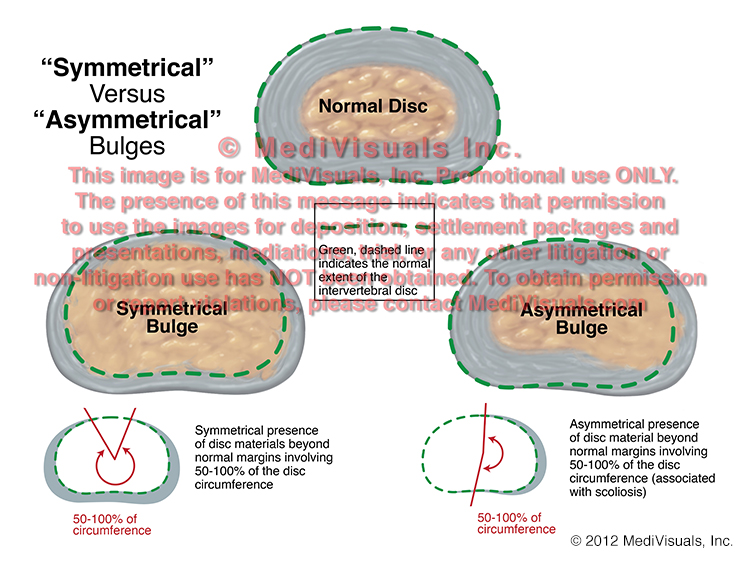
In the image below, a normal disc is shown in comparison to the two types of intervertebral disc injuries covered in this article: "Bulges" and "Herniations". Disc "Bulges", in general, are defined by the presence of disc material beyond the normal margins around at least 50% of the disc's circumference. A "Herniation" is defined as displacement of disc material beyond the limits of the intervertebral disc space that extends less than 50% around the circumference of the disc. The displacement material can consist of the nucleus, the annulus, or parts of both. This is significant in personal injury litigation because the defense often places a great deal of emphasis on whether disc pathology is defined as a "bulge" or "herniation" when determining the severity of an injury. However, a "bulge" can actually impinge nerve roots or the spinal cord to a more severe degree than a "herniation".


The next image compares the normal disc to two different types of disc "Bulges". A "Bulge" is defined as "Symmetrical" when the right and left sides of the herniation more or less mirror each other.  A bulge is "Asymmetrical" when the bulge is more severe on one side when compared to the other.


Finally, the below image shows a normal disc as compared to two types of "Herniations". A "Broad-Based" herniation is defined as disc material extending beyond its normal limits in an area between 25 and 50% of the disc's circumference.  A "Focal" herniation is one involving extension of disc material beyond its normal limits in less than 25% of its circumference.


Annular Tears and Fissures (Intervertebral Disc Pathology, Part 2 of 3)

 By: Robert Shepherd MS, Certified Medical Illustrator, President & CEO, MediVisuals Inc.

This blog is a follow-up referencing language and labels used by health professionals to describe various types of intervertebral disc pathology as defined by a 1995 joint undertaking by representatives from the North American Spine Society, American Society of Spine Radiology and American Society of Neuroradiology.  As a result of their efforts, a more uniform and widely accepted use of nomenclature to define intervertebral disc pathology was developed and published in "Nomenclature and Classification of Lumbar Disc Pathology".

A previous blog dealt with "Symmetrical" and "Asymmetrical" disc "bulge" and "Broad-based" v. "Focal" Herniations.  The first disc pathology term discussed in this blog is "Annular Tear".  This is essentially synonymous with "Annular Fissure," with perhaps "Fissure" being preferable over "Tear" because "Tear" may imply that the pathology was the result of some sort of traumatic event, and this specific pathology can occur without necessarily being the result of trauma.  Annular Tears/Fissures, as seen in the below figure, can occur without fitting the definition of a "Herniation" (disc material extruding beyond its normal boundaries).  As seen in the below illustrations, the fibers of the annulus can be torn with nucleus protruding into the annulus but without the annulus or nucleus extending beyond the bordering vertebral bodies.  By contrast, when "Anular Tears/Fissures" result in disc material extending beyond its normal boundaries, the disc pathology is typically referred to simply as a "Herniation" without a reference to the presence of an Annular Tear/Fissure."


Another blog will be coming soon regarding disc "Protrusions" and "Extrusions".

© 2012 MediVisuals, Inc.

Protrusions Versus Extrusions (Intervertebral Disc Pathology, Part 3 of 3)

 By: Robert Shepherd MS, Certified Medical Illustrator, President & CEO, MediVisuals Inc.


This blog is the third in a series referencing language and labels used by health professionals to describe intervertebral disc pathology as defined by a 1995 joint undertaking by representatives from the North American Spine Society, the American Society of Spine Radiology and the American Society of Neuroradiology.  As a result of their efforts, a more uniform and widely accepted use of nomenclature to define intervertebral disc pathology was developed and published in "Nomenclature and Classification of Lumbar Disc Pathology".

The first blog in the series dealt with "Bulges" v. "Herniations", "Symmetrical" and "Asymmetrical" disc bulges and "Broad-based" v. "Focal" herniations.  The second blog addressed "Anular Tears and Fissures".  This blog addresses the use of "Protrusion" and "Extrusion" to describe intervertebral disc herniations.

"Protrusion" and "Extrusion" are essentially used to further classify types of disc herniations. The term "Protrusion" refers to a disc herniation in which the portion of disc material that is outside the normal confines of the disc space is equal to or less than its aperture where the disc material extrudes from the parent disc.   Examples of disc "Protrusions" and "Extrusions" are shown in the below images. 

The image to the left shows a disc "Protrusion".  Note how the superior and inferior dimensions of the disc material that protrudes from its normal confines (highlighted by the arrow on the left) is not as great as the area where the protruding disc material actually exits its normal confines and boundaries (represented by the arrow on the right in the image).  By comparison, the two illustrations to the right show two different disc "Extrusions".  Note how the dimensions of the protruding disc material are greater than the point where it exits its normal confines. 

It is important to appreciate that disc "Protrusions" and "Extrusions" are terms that may be used to further describe "Broad-based" or "Focal" herniations.  For example, the disc pathology referred to in the above illustration as a "Protrusion" could also be "Broad-Based," if it extends between 25 and 50% of the distance around the circumference of the vertebral body.  Similarly, the disc pathology shown in the illustrations referred to as "Extrusions" could also be referred to as "Focal" if extending less than 25% of the distance around the circumference of the vertebral body (see blog from 08/24/11 for further clarification between "Broad-based" and "Focal" herniations).
© 2012 MediVisuals, Inc.

Disc-Osteophyte Complex Explained

 By: Robert Shepherd MS, Certified Medical Illustrator, President & CEO, MediVisuals Inc.

Individuals who develop new or suddenly worsening symptoms consistent with nerve root or spinal cord impingement following a traumatic event are sometimes diagnosed with “disc-osteophyte complexes”. The term “disc-osteophyte complex” generally refers to abnormal extension of intervertebral disc material that accompanies immediately adjacent osteophyte formation at the vertebral body margin (see the below figure). It is important to note (as shown in the illustrations) that the disc almost always extends further than the osteophytes into the neural foramen or spinal canal to irritate or impinge upon nerve roots or the spinal cord.


Occasionally, individuals who are evaluated shortly after a traumatic event are found to have disc-osteophyte complexes. Because a minimum of several weeks is required for osteophytes to form as a result of a traumatic event, defendant insurance companies may argue that the presence of osteophytes so soon after the traumatic event in question may prove that the plaintiff’s injuries preexisted the traumatic event. Since it is the disc pathology extending beyond the osteophytes that is the actual cause of the nerve root or spinal cord irritation and inflammation, the defense’s arguments are not valid. As shown in the illustrations below, the sequence of events that typically takes place in these cases is that the plaintiff had minimally symptomatic or asymptomatic disc osteophytes prior to the traumatic event in question. During the traumatic event, the disc sustains trauma that results in worsening of the disc pathology while the osteophyte portion of the osteophyte/disc complex remains essentially unchanged. This worsening of the disc pathology in turn results in new or increased irritation or impingement of the neural elements.


© 2012 MediVisuals, Inc


Thursday, June 14, 2012

Medical Legal Illustration and Animation: Cross Sections

 By: Robert Shepherd MS, Certified Medical Illustrator, President & CEO, MediVisuals Inc.

Long before science had advanced to allow imaging of the body in sectional views by computed tomography (CT) and magnetic resonance imaging (MRI), medical illustrators were illustrating the body in sectional views because these views are the best way to appreciate some anatomical relationships. 

Medical illustrators, physicians, and others who have studied anatomy are familiar with sectional views of the body and appreciate the value of these views in explaining the relationship of anatomical structures.  However, accomplished and well respected jury consultants and non-medical illustrator legal graphics experts have expressed concerns that sectional views may be difficult for some jury members to understand.  These individuals' opinions are valuable to those of us in the legal graphics business, and I agree with their opinions that, when other views can communicate a particular relationship message equally as well or better, sectional views should be avoided. I also believe most of these experts will agree that there are times and places in which sectional views of anatomy are the best way to appreciate some anatomical relationships.  Granted, there have been times when we have been working on specific cases and experts have insisted that sectional views be absolutely and unconditionally avoided.  Unfortunately, in these situations the experts were unable to suggest a more effective view to communicate the relevant anatomical relationships (at least in a way that was practical in terms of time and expense). That being the case, sometimes the sectional views were used despite the input of the experts, and at other times, the relationships of the structures had to be explained without the benefits of graphics.
A way to perhaps explain how sectional views help decision makers appreciate relevant anatomical and pathological relationships is to compare them to aerial views or photographs of the scene of a collision.  Space is defined in three planes.  Only two of these planes can effectively be demonstrated in a two-dimensional rendering.  For example, aerial views have long been used to help explain the positions of vehicles and structures that simply can't be appreciate from "street views".   When viewing the scene of a collision from a "street view", one can appreciate vertical and horizontal distances, but not depth; distances close to and far from the viewer's perspective are very difficult to appreciate (see the below figures).  By comparison, when viewing an operative site through a "surgeon's view", vertical and horizontal distances can be appreciated, but the depth of the incision and the relationships of the various structures within and around the incision are very difficult or impossible to appreciate.


The "aerial view" of the collision scene allows the viewer to appreciate distances in two geographical planes as well (distances right and left, and toward and away from the "street view," but the ability to appreciate up and down is lost).  Also, the locations of relevant structures or vehicles that may have been obstructed by nearby structures (such as buildings trees, signs, or other vehicles) can now be appreciated.  Similarly, a sectional view of anatomy can help decision makers appreciate depth relationships of structures.  Or, a sectional view of a step in a surgical illustration can allow the viewer to appreciate the depth of the surgery as well as the additional structures that may have been injured (or at risk of injury) during the invasive procedure.  These specific depth  relationships could not be appreciated from the "surgeon's view" of the same surgery shown in the above illustration.


Exhibits developed to help explain the invasive nature of a surgery and the disruption of the soft tissues during operative procedures are critical.  For that reason, sectional views are critical in aiding a testifying physician to explain these issues.  For example, the exhibit panel that demonstrates an anterior cervical discectomy and fusion (ACDF) that does not include a cross-section through the neck fails to emphasize the depth of the incision and disruption of tissues (essentially all the way to the center of the neck).  This depth simply cannot be appreciated in a "surgeon's view".
In order to appreciate cross-sections, orientation views that show the level and direction of the section are helpful (see below), or when time, budget, and presentation format (digital as opposed to a physical panel) allow, a short animation showing the sectional view actually coming out of the orientation view such as MediVisuals' "Scan SelectorTM" can be used.