MediVisuals

Scroll down to view blog archive

Scroll down to view blog archive

Tuesday, November 27, 2012

Introducing . . . P.I. Exhibits NOW

 By: Robert Shepherd MS, Certified Medical Illustrator, President & CEO, MediVisuals Inc.


P.I. Exhibits NOW  is the newest innovative tool developed specifically for the Personal Injury Attorney. The website is a comprehensive collection of medical exhibits available for purchase and immediate download to a tablet, smartphone, or laptop.

P.I. Exhibits NOW  involves three simple steps:  Search, Purchase, and Download (see below). The site allows for "spur-of-the-moment" availability in depositions, settlement hearings, mediations, or trials. The collection consists of over 400 exhibits demonstrating many of the injuries and surgeries that personal injury attorneys routinely address, and which best lend themselves to "generic" or "typical" representations. 


The effectiveness of visuals is recognized throughout society in such common phrases as “A picture is worth a thousand words”, and “Seeing is believing.” Additionally, numerous manuscripts refer to studies substantiating that recall is greatly increased when a verbal message is supported by visual images. These studies have typically shown that, after varying periods of time, information presented vocally and supported by visuals was recalled at a significantly higher rate than the same message delivered by voice alone.


Visit P.I. Exhibits NOW.  Images purchased are projection resolution and are available for $245 each, a savings of $50 compared to MediVisuals.com. Click here or go to http://store.medivisuals.com/.


References
DeBoth, C. J., & Dominowski, R. L. Individual differences in learning: Visual versus
auditory presentation. Journal of Educational Psychology, 1978; Aug 70 (4): 498-503.
McCall, J., & Rae, G. Relative efficiency of visual, auditory and combined modes of
presentation in learning of paired-associates. Perceptual and Motor Skills, 1974; June (38): 955-958.
Multimodal Learning Through Media:  What the Research Says. Cisco Web site. http://www.cisco.com/web/strategy/docs/education/Multimodal-Learning-Through-Media.pdfAccessed August 19, 2008.

Wednesday, August 8, 2012

Pneumothorax, Hemothorax, Pulmonary contusions - The Overlooked Life-Threatening Injuries

 By: Robert Shepherd MS, Certified Medical Illustrator, President & CEO, MediVisuals Inc.

When developing medical demonstrative aids for demand packages, mediations, depositions, or trial, the personal injury attorney typically expresses a desire to emphasize the broken bones but may casually disregard injuries to the lungs such as pneumothorax, hemothorax, or pulmonary contusions.  When a plaintiff suffers both broken bones and these pulmonary injuries, they often require a prolonged hospitalization -- not because of the broken bones, but because of respiratory compromise related to these lung injuries that may require intubation, chest tubes, or tracheostomies and may advance to pneumonia, adult respiratory distress syndrome, or even death.

When applicable and practical, demonstrative aids addressing these pulmonary injuries can be very helpful in emphasizing the life-threatening nature of the injuries and in opening the doorway for testifying physicians to explain permanent lung pathology that may have developed as a result of these injuries.
Negative pressure has to be present between the lungs and chest wall in order for the lungs to inflate during inspiration.  A hemothorax (accumulation of blood between the lungs and chest wall) typically is a result of injury to internal tissues or blood vessels that bleed into the chest cavity.  A pneumothorax (accumulation of air between the lungs and chest wall) typically results from a perforation of the lung or the chest wall that allows air to enter the space between the lungs and chest.  A hemopneumothorax is an accumulation of both air and blood between the lungs and chest wall.  With blood and/or air in this space, the ability of the injured person to breath can be significantly impaired resulting in the need for intubation, tracheostomy, chest tubes, or at least prolonged hospitalization.

A pulmonary contusion (also referred to as pulmonary hematoma), as well as atelectasis, can also result in respiratory compromise, prolonged hospitalizations, intubation, and multiple complications as previously discussed.  Pulmonary contusions or hematomas occur when blood accumulates within the lung itself -- either within the lung tissue or within the alveoli (the air pockets where oxygen is absorbed into the blood).  Atelectasis is a more generalized condition that refers to the failure of the alveoli to be adequately expanded and allow oxygen and carbon dioxide to exchange.  Following trauma, the cause of atelectasis (post-traumatic atelectasis) can be the result of several issues such as the patient's inability to adequately expand their chest during inspiration (due to chest trauma or as a side effect of pain medications).  Another common cause of post-traumatic atelectasis is compression of the lung tissue by injured and swollen abdominal organs or collections of abdominal blood that push upward on the diaphragm and prevent the lungs from adequately inflating during inspiration.

© MediVisuals, Inc. - Permission to use any image (or parts thereof) posted on this blog in depositions, demand packages, settlement hearings, mediation, trial, and/or any other litigation or non-litigation use can be obtained by contacting MediVisuals at www.medivisuals.com – otherwise copyright laws prohibit their use for those or other purposes.

Thursday, July 26, 2012

"Minimally Invasive" Spine Treatments: Discography, Injections & Ablation

 By: Robert Shepherd MS, Certified Medical Illustrator, President & CEO, MediVisuals Inc.

On occasion, it is uncertain exactly which intervertebral disc may be causing a plaintiff’s pain.  Physicians may conduct a discography study prior to surgery in these cases.


This procedure involves advancing needles into the discs in question and injecting contrast material that serves two purposes:  (1) The contrast material makes it possible to better analyze the exact defects (if any) in the discs when X-rays or CTs are taken after the administration of the contrast material. (2) The contrast material also increases the pressure within the disc causing it to expand and subsequently compress the nearby nerve roots.  If     the pain corresponds to the patient’s normal pain, then that disc is determined as the     “problem disc”.


When pain is thought to be associated with irritation or inflammation of the nerve root(s) or surrounding tissues, epidural injections can be performed. As shown in the animation below, epidural injections involve administering anesthetics and steroids around the nerve root(s).  These injections can be used as a diagnostic tool as well as a treatment.


If the epidural injections are ineffective, the cause of the pain may not be related to the nerve root(s).  If the injections are effective, it confirms the nerve root(s) as the source of the pain. Repeated treatments may resolve the pain; if not, more aggressive treatments may be necessary.
Sometimes the facet joints themselves can be the source of pain. One of the terms used to refer to this condition is “facet arthropathy”.  This condition results from the breakdown of the normal, healthy joint spaces. With the breakdown of these articular surfaces, the joints become painful with each movement.


Each facet joint is supplied by a small dorsal branch of the adjacent nerve root [see illustration below].  Injections in or around the facet can again serve to determine the painful facet(s) and help resolve the pain.


If facet joint injections are unsuccessful at resolving the pain, the nerves to the facets can be destroyed by a procedure known as ablation. Ablation involves advancing a needle adjacent to the facet nerve and destroying it, thereby eliminating the pain.


Understanding the Brachial Plexus Injury: Part 2 (Shoulder Dystocia)

By: Trisha Kreibich, MS, Medical Illustrator/Consultant, and Robert Shepherd MS, Certified Medical Illustrator, President & CEO, MediVisuals Inc.

This article is a continuation of a two part article on brachial plexus injury. Part 1 covered brachial plexus injuries in adults caused by a traumatic event, such as a motor vehicle collision. Part 2 will address brachial plexus injury in infants during delivery, which is also known as Shoulder Dystocia or Erb's palsy.

To review, the brachial plexus innervates the arm and is formed by several of the cervical nerve roots and the T1 nerve root. [see illustration below]


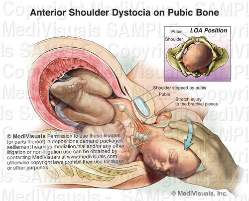
During delivery, the infant's passage through the birth canal can be stopped by the impaction of its shoulder(s) against the mother's pubic bone, sacral promontory, or both. [see illustration below]


Injury to the brachial plexus may sometimes occur because of unusually powerful uterine contractions or rapid fetal descent; however, brachial plexus injury may also result from improper obstetrician interaction. When excessive downward traction on the head of an infant with shoulder dystocia is applied (a violation of the standard of care), the delicate brachial plexus is stretched and injured. [shown below] The same may also occur when vacuum extraction is used in this situation.


As in adult brachial plexus injuries, the earlier the injury is identified and treated, the better chances are for recovery and preventing permanent damage. However, this is also dependent on the severity of the initial injury. Sometimes, an overgrowth of nerve cells (neuroma) can form around the injured nerves. When this occurs, surgical intervention and nerve grafting may be needed. [see illustration below]


There are some delivery techniques that can be used to prevent brachial plexus injury in infants with shoulder dystocia. As the illustration below demonstrates, the mother is moved to the McRoberts position, where her legs are hyperflexed to the abdomen, typically resulting in an increase of the outlet. This is often used in conjuction with an episiotomy, a cut made in the perineal body (tissue between the vagina and anus) before delivery, to enlarge the outlet and allow the obstetrician more room to perform maneuvers.


If no progress is made, moderate suprapubic pressure may be applied to free the impacted shoulder. The Wood's screw maneuver [shown below] may be used as well. This maneuver involves the obstetrician rotating the infant's anterior or posterior shoulder, and in turn the body, like a screw, freeing the impacted shoulder.


The following animation was developed to show how brachial plexus injuries can occur in a shoulder dystocia case, along with a few of the above mentioned accepted procedures that can be performed to help prevent this injury.


Reference:
Gabbe, S.G., Niebyl, J. R., & Simpson, J.L. "Obstetrics: Normal & Problem Pregnancies." 3rd ed. Philadelphia: Churchill Livingstone, 1996. 374-375, 490-494. Print.

Understanding the Brachial Plexus Injury: Part 1

 By: Robert Shepherd MS, Certified Medical Illustrator, President & CEO, MediVisuals Inc.

Injuries to the brachial plexus can often take place from trauma similar to that which causes cervical spine injuries and can also manifest similar symptoms. As shown in the illustration below, the brachial plexus is formed by several of the cervical nerve roots and the T1 nerve root.


Sometimes injury to the brachial plexus can be the direct result of excessive stretch during a traumatic event. For example, in a motor vehicle collision with a violent side impact the nerves on the contralateral side of the impact (and resulting flexion) may be stretched and/or torn. [see illustration below]


Injury to the surrounding muscles can indirectly cause injury to the brachial plexus, as well. The brachial plexus runs between the anterior and middle scalene muscles, which connect the cervical spine and first rib. In a motor vehicle collision, hyperextension of the neck may excessively stretch these muscles, as demonstrated in the illustration below.


When these muscles are stretched, the resulting swelling or spasm can result in symptoms consistent with cervical nerve root injury even though the cervical nerve roots or brachial plexus themselves may not be directly injured. [see illustration below]


Thoracic outlet syndrome is another mechanism by which the brachial plexus can be injured. The illustration below shows that the axillary sheath, which contains the nerves from the brachial plexus and the axillary vein and artery, passes through the opening created between the clavicle and first rib.


Thoracic outlet syndrome can occur either by (1) the elevation of the first rib due to spasm of the scalenes or (2) the loss of innervation to the trapezius and/or other muscles that insert on the clavicle or scapula, causing the shoulder to droop [see illustration below]. By either mechanism, the opening between the clavicle and first rib is closed and the nerves and blood vessels that travel through the thoracic outlet become compressed.


As one can see in the illustration below, an injury to the brachial plexus affects the motor and sensory function of the arm. The earlier the injury is identified and treated, the better chances are for recovery and preventing permanent damage. However, this is also dependent on the severity of the initial injury. Sometimes surgical intervention is needed to address torn/ruptured nerves and excessive scarring.


This is the first of a two part article. Please check back to read Part 2, which discusses brachial plexus injury in infants during delivery (Shoulder dystocia). You can also sign up for email alerts, which announce when another article has been posted.


Vestibular Nerve Injury: Why it could be important to your TBI case

 By: Robert Shepherd MS, Certified Medical Illustrator, President & CEO, MediVisuals Inc.

Balance and dizziness are often associated with traumatic brain injuries, although the specific cause of these problems is often difficult to explain. Sometimes the injuries may be to the inner ear organs. Other times the injuries may be to the vestibular nerve. When the injury is to the vestibular nerve, the mechanism of injury is similar to injuries to the olfactory nerve resulting in disturbances in smell.

The exhibit shown below demonstrates the mechanism of injury. As the brain stem and skull move in different directions during a violent impact, stretch injuries to the vestibular nerve can occur. This type of injury is especially significant when supporting arguments of brain injuries occurring as a result of traumatic forces to the head. If forces were significant enough to damage the vestibular nerve, the forces were likely sufficient to cause shear or traumatic axonal injury, as well.


For more information on mild and severe traumatic brain injury, please visit: http://www.medivisuals.com/traumatic-brain-injury.aspx For more information on the featured exhibit, please visit: http://www.medivisuals.com/vestibular-nerve-injury-mvi82010-01x.aspx

Discogenic Pain - Back Pain and Radiculopathy Without Evidence of Significant Disc Injury

 By: Robert Shepherd MS, Certified Medical Illustrator, President & CEO, MediVisuals Inc.

In determining if a person's pain may be related to some sort of intervertebral disc pathology, a great deal of emphasis is placed upon imaging studies showing evidence of mechanical compression of a nerve root by abnormal posterior displacement of a disc (i.e. bulge, protrusion, herniation, etc.) as portrayed in the below illustration.


In cases where clear mechanical compression of the nerve roots is not shown in imaging studies, some are quick to argue that any pain emanating from the area is either exaggerated or entirely contrived. However, a person can experience pain consistent with mechanical compression of a nerve root without having any significant disc pathology. This is because the spine is encircled with a meshwork of nerves that are much too small to be seen on CT or MRI (see the below figure). The sinuvertebral nerves surround and penetrate the intervertebral discs.

When injuries to a disc are more subtle, the sinuvertebral nerves may detect the injuries and send pain signals to the brain where they are interpreted as pain (see figure). The pain may be limited to the area of the back, or a pain perception phenomenon know as "pain referral" (confusion of the origin of pain signals by the brain) may result in the person experiencing very real pain consistent with radicular pain from mechanical nerve root compression by a severely herniated disk.

Another common cause of pain consistent with nerve root mechanical compression is chemical irritation or inflammation of the nerve root. Chemical irritation of a nerve root often results from the release of chemicals following a more subtle disc injury (see the below figure). These chemicals irritate and inflame the nerve root and surrounding tissues, resulting in the perception of pain consistent with an injury to the disc and mechanical compression of the nerve root. Even after resolution of chemical irritation and inflammation, scar tissue may develop that binds the nerve root (often undetectable on CT or MRI). This scarring can cause permanent debilitating pain that may require surgical intervention.


© MediVisuals, Inc. - Permission to use any image (or parts thereof) posted on this blog in depositions, demand packages, settlement hearings, mediation, trial, and/or any other litigation or non-litigation use can be obtained by contacting MediVisuals at www.medivisuals.com – otherwise copyright laws prohibit their use for those or other purposes. 

Soft Tissue Injury - Cervical and Lumbar Strain

 By: Robert Shepherd MS, Certified Medical Illustrator, President & CEO, MediVisuals Inc.

Injuries to the spinal and paraspinal ligaments and muscles can result from violent side-to-side motions or by violent excessive flexion and extension. The illustration below shows the major ligaments of the neck (anterior longitudinal and interspinal ligaments) in hyperflexion and hyperextension, which can be injured grossly or microscopically.


The series of three illustrations in the bottom right corner (which is shown in more detail below), show a close-up view of the spinal anatomy in 1.) the Normal condition, 2.) during Excessive Stretching and 3.) After Healing. In the Normal condition, one can appreciate the close relationship between the muscles, nerves and blood vessels. During Excessive Stretch, small tears occur, which causes bleeding in the muscle fibers. After Healing, scar tissue and inflammation entrap blood vessels and nerves resulting in a permanent state of compromised, painful movement.


The same is true for the lower lumbosacral spine and pelvic regions. In the illustration below, the spinal nerves and their posterior branches are seen in close approximation to the ligaments and joint capsules, which are often involved in the injury. During hyperflexion of the lumbar spine, transient bulging of the intervertebral discs can occur. 


The series of illustrations in the lower right corner of the above image, show the normal lumbosacral and pelvic muscles and tendon fibers, which insert on the bones near the associated posterior spinal nerve branches. During Excessive Stretch, a segmental artery and its branches may be involved in hemorrhaging, scarring and occlusion. After Healing, scar tissue and adhesions form, entrapping nerves and blood vessels, causing chronic pain.

In these next illustrations, muscle is shown in sequentially higher magnifications, which can be used to explain excessive stretch injuries in any area of the body.  In the Normal series, the bottom illustration depicts nerves and small body vessels intertwined in muscle with its tendinous attachment to the bone.  The middle illustration shows a magnified view of an individual muscle fiber and the top illustration depicts the relationship of the microscopic myofilaments (actin and myosin) in their normal relaxed position.


In the Excessive Stretch series, the bottom illustration shows the muscle, tendon, blood vessels and nerves as they are excessively stretched. Small hemorrhages are seen escaping from the stretched and torn blood vessels. In the middle illustration, blood is shown escaping into surrounding spaces, reducing oxygen exchange to the muscle and irritating the delicate structures of the muscle fiber. The top illustration depicts the myofilaments, showing the actin and myosin fibers torn and stretched past the point of normal interdigitation.


In the After Healing series, the bottom illustration shows the irregular outline of the scarred and inflammed muscle fiber with small adhesions seen between the blood vessels, nerves and muscle fibers. The middle illustration shows scar tissue and inflammation occluding blood vessels and adhering the delicate structures of the muscle fibers together, limiting motion and causing chronic pain. Lastly, the top illustration depicts the damaged myofilaments. Their normally well-organized, interdigitating arrangement is left destroyed, limiting muscle movement at the most basic level.


Monday, July 16, 2012

Disc Herniation and Other Disc Injuries


 By: Robert Shepherd MS, Certified Medical Illustrator, President & CEO, MediVisuals Inc.

The term “degenerated disc” is generally used to describe a disc in the early degenerative process. It is the beginning of a progressive break down of the disc. This condition can be initiated or accelerated by a traumatic event.


A disc bulge is a more advanced collapse of the disc to the point that the disc expands beyond its normal contour. It may or may not impinge on the neural structures within the spinal canal or neural foramina. Similarly, a disc bulge can be the immediate or delayed result of a traumatic event, or a traumatic event may exacerbate a preexisting, stable disc bulge.

A subligamentous herniation is one in which nucleus pulposus has extended through the annulus fibrosus, but has not gone through the posterior longitudinal ligament.


The term “herniation” is generally used when the nucleus has completely extruded through the annulus fibrosus and posterior longitudinal ligament.

However, it does not matter what the disc pathology is labeled, if it impinges upon, or irritates the neural components, it is a significant injury that will likely require some type of invasive procedure to correct.


The disc can either be injured by an immediate tear of the annulus fibrosus and extrusion of the nucleus pulposus during a traumatic event, or they can be the result of a much more gradual process. In order to understand the gradual breakdown of the disc, one must first understand a little of the physiology of a disc. The inner disc relies on exchange of fluid, nutrients and oxygen through the end plate of the adjacent vertebral bodies.
During a traumatic event, the endplate may become injured resulting in interference with that exchange.


As a result of the inability of the disc to obtain the fluid, nutrients, and oxygen it needs, the disc gradually begins to break down - becoming a degenerated disc, followed by a bulging disc, and eventually to a herniated disc. The amount of time involved with the process of the disc breakdown is related to the severity of the initial disc injury.


Therefore, if a disc injury is not evident until weeks or months after a traumatic event, it does not mean the injury was not a direct result of the traumatic event.


© MediVisuals, Inc. - Permission to use any image (or parts thereof) posted on this blog in depositions, demand packages, settlement hearings, mediation, trial, and/or any other litigation or non-litigation use can be obtained by contacting MediVisuals at www.medivisuals.com – otherwise copyright laws prohibit their use for those or other purposes.


Thursday, July 5, 2012

Breaking Down Traumatic Arthritis

 By: Robert Shepherd MS, Certified Medical Illustrator, President & CEO, MediVisuals Inc.

Fractures can result in several long term or permanent complications that can necessitate additional surgical procedures. One of the most common long term debilitating complications is traumatic arthritis.Traumatic arthritis can affect almost any moveable joint in the body. To explain traumatic arthritis more in depth, we will be focusing on the tibiotalar (ankle) joint, as shown in the illustration below.
As like most moveable joints, the tibiotalar joint consists of smooth articular bone covered by thick, shock-absorbing articular cartilage.
During trauma, the joint surfaces can be driven together resulting in injuries to the cartilage and microfractures of the articular surface (even without obvious intrarticular fracture).
The bone and cartilage then undergo changes that result in the progressive breakdown of the joint. As the process advances, the joint becomes painful. In most cases, the only treatment options are joint replacement or fusion.
The acromioclavicular (AC) joint also frequently falls victim to traumatic arthritis. As the AC joint enlarges (hypertrophy) it impinges on the rotator cuff, which is referred to as subacromial impingement. This can cause irritation or tearing of the rotator cuff and is most often treated byAC joint resection and subacromial decompression.